GI Endoscopy · 6 min read
Griffith's Point in 2026: Splenic-Flexure Watershed and Drummond
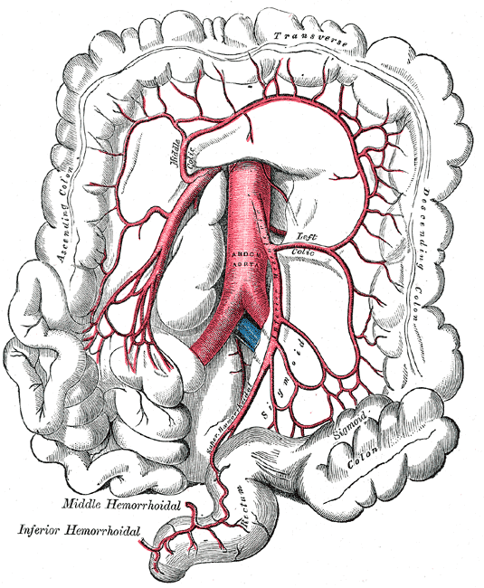
The marginal artery of Drummond is the colon's collateral arcade. It is often weak or absent at the splenic flexure. That weak point is Griffith's point.
Griffith's point is the watershed at the splenic flexure between middle-colic (SMA) and left-colic (IMA) flow. The marginal artery of Drummond is supposed to bridge those territories. At this bend the arcade is often narrow or missing, which is why systemic hypotension paints the splenic flexure first. The arcade is the collateral. Griffith's point is where that collateral fails.
Experienced teaching points
Clinical Pearls
- Drummond is the arcade along the mesenteric border. Griffith's point is the weak spot in that arcade at the splenic flexure. Do not retitle one as the other.
- About 5% of people have a functionally absent marginal artery at the flexure. Those are the hypotensive left-colon infarcts that surprise a normal IMA origin.
- The arc of Riolan is a more proximal, meandering SMA-IMA vessel. It is not Drummond. A hypertrophied Riolan on angiography is a chronic SMA or IMA occlusion until proven otherwise.
- Typical medical colon ischemia maps to this watershed. Rectosigmoid ischemia after IMA ligation maps to Sudeck's point.
- Anatomy does not replace CTA when the right colon is the only segment. That is SMA territory, not Griffith.
Clinical Bottom Line
| Question | 2026 practical answer |
|---|---|
| What is Griffith's point? | The watershed at the splenic flexure between the middle colic artery (SMA) and the left colic artery (IMA). Griffiths described the critical anastomosis in 1956. Meyers put it on the radiology map in 1976. |
| What is Drummond? | The marginal artery running 1 to 3 cm from the mesenteric border, built from ileocolic, right colic, middle colic, left colic, and sigmoid arcades. It is the usual SMA-IMA collateral. It is often tenuous exactly at Griffith's point. |
| Why does hypotension hit here? | Terminal branches, lowest collateral reserve. A drop in systemic pressure fails this bend before a well-arcaded transverse or sigmoid colon. That is ordinary nonocclusive colon ischemia. |
| Arc of Riolan? | A more central meandering mesenteric artery, not the paracolic Drummond arcade. A fat Riolan is a clue to chronic proximal SMA or IMA occlusion, not a Griffith diagnosis. |
| Clinical page | Colon ischemia for CT, colonoscopy, IRCI, and surgery. Sudeck's point for the rectosigmoid IMA story. |
Where is Griffith's point?
John Griffiths mapped the blood supply of the distal colon in 1956 and called out a critical anastomosis at the splenic flexure. Meyers's 1976 AJR paper is the one most radiologists still quote: Griffith's point as the SMA-IMA watershed whose failure explains a subset of colon ischemia. The middle colic (SMA) and left colic (IMA) are supposed to meet here through Drummond. Sometimes they barely connect.
Drummond is the arcade. Griffith's point is the weak spot in that arcade. Teach both names. Do not use one as a nickname for the other.
What is the marginal artery of Drummond?
Drummond is a continuous (in textbooks) paracolic arcade. Ileocolic, right colic, middle colic, left colic, and sigmoid branches anastomose 1 to 3 cm off the bowel wall and can reverse flow when one parent trunk is gone. That is how a chronic IMA occlusion can be silent, and how a high IMA ligation can still leave a pink descending colon.
The textbook is a best case. At the splenic flexure the connecting vessel is often small, interrupted, or absent. Older anatomic series put a functionally missing flexure anastomosis in the neighborhood of 5%. Those patients have a Griffith's point in the original sense: a true watershed with no backup. They are the hypotensive left-colon injuries with a widely patent SMA and IMA origin on CTA. The pipes are open. The last mile is not.
When you ligate the IMA, you are asking Drummond to carry the left colon on middle-colic pressure. If Griffith's point is the missing link, the descending colon is the next ischemic segment, not just the rectosigmoid. Check the cut edge. Do not trust the drawing.
Why does hypotension hit the splenic flexure?
Nonocclusive colon ischemia is a mucosal flow problem. The watershed with the poorest collateral wins. Empirically that is the left colon, often the flexure and proximal descending colon. Brandt's 313-case map still has the left colon as the most common segment. ACG 2015 keeps Griffith's point in the teaching diagram for that reason.
This is the medical patient: shock, dialysis, heart failure, a long run, a vasoconstrictor, a constipation cocktail. The IMA origin is usually open. Angiography after the fact is often normal. That is why ACG does not want a mesenteric angiogram as the first test for ordinary left-sided colon ischemia. Read the clinical page for CT, timing of colonoscopy, and who goes to surgery.
Isolated right-colon ischemia is not a Griffith problem. The right colon lives on the SMA. Pain without bleeding plus a thick right colon is AMI-adjacent until CTA says otherwise. Do not invoke a splenic-flexure watershed to skip the angiographic phase.
What about the arc of Riolan?
The arc of Riolan (meandering mesenteric artery) is a more proximal SMA-IMA connection, usually between middle colic and left colic nearer the root of the mesentery. It is not Drummond. A hypertrophied Riolan on CTA is a chronic occlusion of SMA or IMA until you prove otherwise. It can save a colon. It can also mark a patient whose next hypotensive hit will not be so polite.
Name the vessels in the report. "Good collaterals" is not a vessel.
Selected references
- Griffiths JD. Surgical anatomy of the blood supply of the distal colon. Ann R Coll Surg Engl. 1956;19:241-256.
- Meyers MA. Griffiths' point: critical anastomosis at the splenic flexure. Significance in ischemia of the colon. AJR Am J Roentgenol. 1976;126:77-94.
- Brandt LJ, Feuerstadt P, Longstreth GF, Boley SJ. ACG Clinical Guideline: Colon Ischemia (CI). Am J Gastroenterol. 2015;110:18-44.
- Brandt LJ, Feuerstadt P, Blaszka MC. Anatomic patterns, patient characteristics, and clinical outcomes in ischemic colitis. Am J Gastroenterol. 2010;105:2245-2252.
- Sudeck's point and colon ischemia.
Last reviewed September 20, 2026. Written for clinicians who were taught Drummond as a continuous safety net, and need the splenic-flexure exception before they interpret a left-colon CT.
Topics
For your teaching file
Save this article as a PDF
Drop your email and we'll open a print-ready version you can save as a PDF — and you'll start getting our weekly GI endoscopy newsletter.