GI Endoscopy · 3 min read
Variceal Hemorrhage in 2026: Triage, Banding, Early TIPS, and Gastric Varix Decisions
A practical update on acute variceal hemorrhage: first-hour orders, endoscopic band ligation, early TIPS selection, gastric varix therapy, and secondary prophylaxis.
Clinical Bottom Line
| Decision Point | Current Answer | Practice Move |
|---|---|---|
| Initial suspected variceal bleed | Resuscitation, vasoactive therapy, antibiotics, restrictive transfusion, and urgent endoscopy still anchor care. | Build the order set before the patient arrives in the endoscopy unit. |
| High-risk esophageal variceal bleeding | Early TIPS should be considered in selected high-risk patients rather than waiting for repeated failure. | Trigger hepatology and interventional radiology early when Child-Pugh C or high-risk Child-Pugh B features are present. |
| Gastric fundal varices | Banding logic does not translate cleanly to fundal varices. | Think cyanoacrylate or EUS-guided therapy where available, plus TIPS or retrograde transvenous obliteration depending on shunt anatomy and liver reserve. |
What Changed in the Last 12 Months
The most useful recent shift is operational rather than conceptual. Current guidance and reviews emphasize earlier risk stratification, noninvasive portal hypertension assessment, and faster escalation for high-risk bleeding rather than treating all variceal hemorrhage as a single endoscopic problem.
The old framing that a red wale mark predicts a catastrophic bleed in the next 48 to 72 hours is too deterministic. Red wale signs matter, but acute management depends on hemodynamics, cirrhosis severity, active bleeding, infection risk, renal function, encephalopathy risk, and whether the varix is esophageal or gastric.
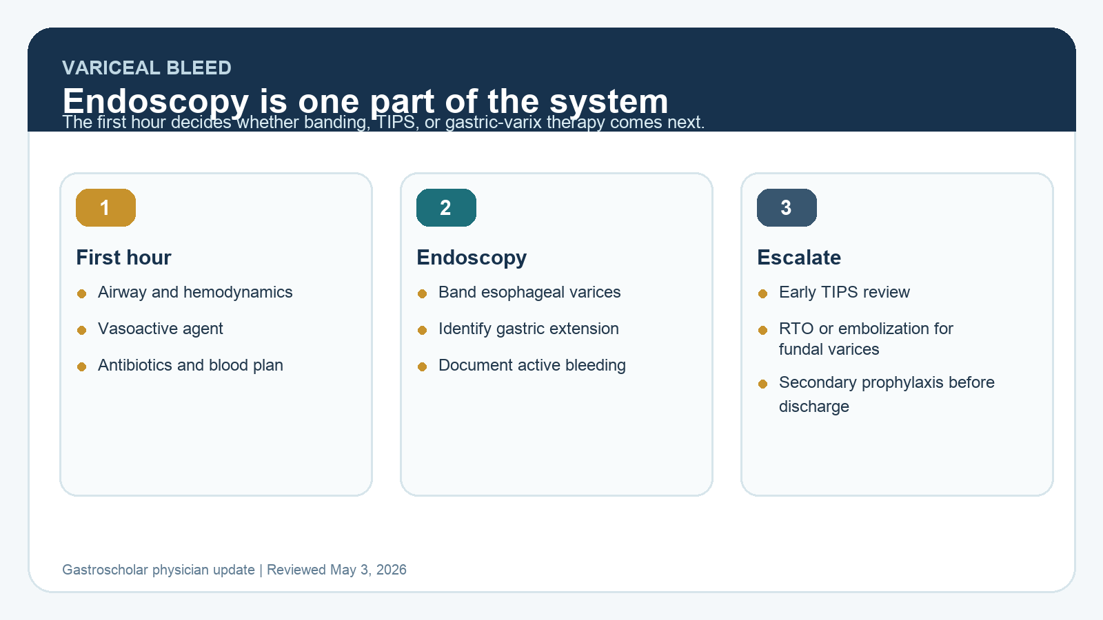
First Hour Management
Variceal hemorrhage is a systems problem. The endoscopist matters, but outcomes depend on what happens before scope insertion.
- Stabilize airway, circulation, mental status, and aspiration risk.
- Start vasoactive therapy promptly when variceal bleeding is suspected, before endoscopy confirms it.
- Give antibiotic prophylaxis in cirrhosis with upper GI bleeding unless a clear contraindication exists.
- Use a restrictive packed red blood cell strategy in most patients, while individualizing for shock, ischemia, ongoing hemorrhage, and massive transfusion needs.
- Correct severe coagulopathy or thrombocytopenia selectively. Do not turn the INR into the main therapeutic target.
Endoscopy: What Still Matters
For esophageal variceal bleeding, endoscopic variceal ligation remains the core endoscopic therapy. The key quality issue is not simply placing bands. It is recognizing active bleeding, clearing the field safely, avoiding over-insufflation, identifying gastric extension, and deciding when endoscopy alone is not enough.
For gastric varices, location changes the plan. GOV1 can often be managed similarly to esophageal varices. GOV2 and isolated gastric fundal varices require a different mental model because embolic and shunt-based therapies may be more important than repeated banding.
When to Escalate Beyond Banding
Early TIPS
Early or preemptive TIPS is not for every bleed. It belongs in the discussion for selected high-risk patients, especially those with severe liver disease or active bleeding despite appropriate initial therapy. The decision is time-sensitive and should involve hepatology and interventional radiology early.
Gastric varix pathways
For gastrofundal varices, AASLD guidance highlights the role of TIPS, variceal embolization, and retrograde transvenous obliteration. The best choice depends on anatomy, spontaneous shunts, portal vein patency, ascites, encephalopathy risk, transplant candidacy, and local expertise.
How to Apply This in Practice
- Create a variceal bleed activation checklist: octreotide or equivalent vasoactive agent, ceftriaxone or local antibiotic pathway, blood bank plan, ICU criteria, airway risk, and endoscopy target time.
- At the first endoscopy, document varix type, active bleeding, stigmata, therapy used, completeness of hemostasis, and whether gastric varices were present.
- Before the patient leaves the unit, decide whether they need early TIPS review, gastric varix imaging, transplant-center discussion, or secondary prophylaxis planning.
- For secondary prophylaxis of esophageal varices, combine nonselective beta-blockade with serial ligation when tolerated and appropriate.
- After RTO-type therapy, remember that esophageal varices can worsen as portal pressures redistribute.
Practice Pitfalls
- Waiting until failed repeat banding before calling interventional radiology in a high-risk bleed.
- Treating gastric fundal varices as if they were distal esophageal columns.
- Over-transfusing stable patients and potentially increasing portal pressure.
- Skipping antibiotics because the bleeding source seems "purely mechanical."
- Discharging without a secondary prophylaxis plan, repeat endoscopy timeline, or hepatology follow-up.
Key Sources
- AASLD portal hypertension bleeding in cirrhosis practice guidance page
- AASLD guidance on TIPS, variceal embolization, and retrograde transvenous obliteration
- 2025 review on endoscopic management of portal hypertension and varices
- 2025 review on diagnosis and management of clinically significant portal hypertension
- 2026 GESA consensus on portal hypertension in cirrhosis
Clinical guidelines summarized by the Gastroscholar Research Team. Last updated: May 3, 2026. This article is intended for physicians and advanced clinicians.
For your teaching file
Save this article as a PDF
Drop your email and we'll open a print-ready version you can save as a PDF — and you'll start getting our weekly GI endoscopy newsletter.